Erbacres Holsteins recognized as the 2026 McKown Master Breeder Award
Erbacres Holsteins recognized as the 2026 McKown Master Breeder Award
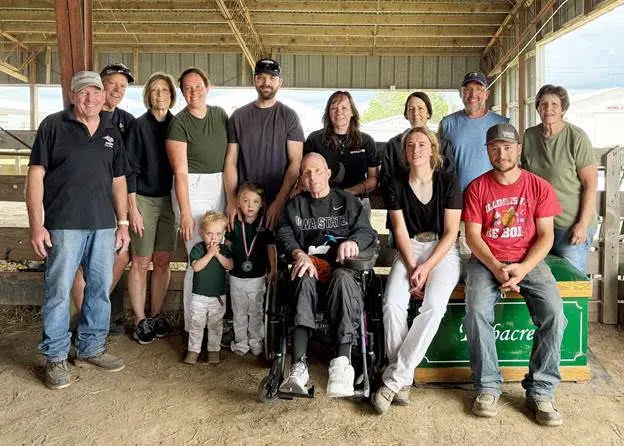
Close family ties and a passion for breeding high-caliber cows are among the most notable traits of Erbacres Holsteins of Lanark, Ill., this year’s recipient of the Robert “Whitey” McKown Master Breeder Award. This award is presented to breeder herds that are known for combining strong genetics, production, longevity, and breed influence. Hallmarks for this award are dedication to the farm and the breed, reflecting on multigenerational commitment and persistence to the industry. These characteristics are key definers for the Erbsen family.
The farm initially began with a small herd of grade Holsteins in 1957 following the marriage of Kenneth and Pauline Erbsen. Rather quickly, the grade Holsteins were dispersed and replaced with registered Holsteins. That led to the debut of the Erbacres prefix. The farm has been completely family owned and operated since those early days and has grown into the perfect family affair.
Kenneth and Pauline raised six children with integrity for the farm and love for the Holstein cow – Paula (Bovre), Kathleen (Opperman), Kevin, John, Carl, and Carla (Mickey). With the growth of the herd, the Erbsen family started milking at a second location in 1975. After Kenneth’s passing in 1994, the Erbsen family dispersed one barn of milk cows, and the oldest son, Kevin, took over managing the second farm along with the breeding decisions for the herd, assisted on the farm by his mother Pauline – until her passing in 2021. Kevin’s brothers John and Carl assist as their off-farm jobs allow.
In February of this year, Kevin faced some unexpected health challenges and had to step back from daily operations. Consequently, the next generation, Kevin’s children – son and daughter, Payton and Delana, along with Kevin’s wife, Wendy, have taken on an expanded role in farm responsibilities and decision making. With Kevin’s health issues, the entire extended Erbsen family also has come together to keep the farm running smoothly.
These days, the Erbsen family crops 700 acres of land and raises 100 head of registered Holsteins. While the Erbacres herd may be small, the cows have proved themselves mighty. The farm has bred 245 Excellent cows, 21 Gold Medal Dams, 25 Dams of Merit, and received the Progressive Breeders Registry award for 28 years. The 42 milking cows average a 112.5% Breed Age Average (BAA) with production records of 21,357 average pounds of milk, 4.3% fat, and a 3.3% protein.
The Erbsen family began showing when Paula, the oldest child, reached nine years of age. That means that the family has been exhibiting at their District Holstein Show – now called the Northern Illinois Show – for 59 years. The family won Grand Champion of their district show in 1974 with Erbacres Marie Hagen Marigold. Marigold went on to the first of nine champions at the Illinois Championship show or Illinois Championship Junior Show, along with numerous reserve champions, All-Illinois, and Reserve All-Illinois winners through both open and junior shows.
Throughout the years, Erbacres has exhibited 25 Illinois Futurity winners, including the 2026 five-year-old Futurity winner, Erbacres Tatoo Cleo EX-93. They also exhibited the Pabst Futurity winner in Wisconsin in 1993 with Erbacres Lancer Cherokee EX-91 GMD DOM. Additionally, Erbacres has been awarded the Premier Breeder banner at the Illinois Championship Show 25 times, along with winning Premier Breeder at other shows including the Illinois State Fair, Wisconsin State Fair, and Wisconsin Spring Show.
The Erbacres prefix has been worn by several show winning cows, but there is one cow that rises to the top of that list: Erbacres Snapple Shakira-ET *RC, scored at EX-97 4E 10*. Shakira was Supreme Champion not once, but twice at World Dairy Expo in 2021 and 2023, as well as Supreme Champion at The Royal Agricultural Winter Fair in 2023.
Additionally, Shakira has several daughters’ gaining traction in the show ring, along with having several sons in stud. Shakira has become a standard bearer for the Holstein breed, embodying genetic excellence and competitive accolades. Additional individuals to note wearing the Erbacres prefix are:
- Erbacres Damion EX-96 who is a former #1 Type sire at Select Sires
- Erbacres Rubens Libby-Red EX-94 who was named All-American Red and White Jr. 3-Year-Old and won the Red and White Futurity at the World Dairy Expo in 2005
- Erbacres Magician, a two-time All-American and Grand Champion bull at the 1992 International Show at World Dairy Expo.
Breeding for type traits has always been emphasized within the breeding decisions at Erbacres. The Erbsen family feels that high type cows last within the operation and are profitable producers for several lactations.
The Erbsen family has been heavily involved within the Illinois Holstein Association, and the younger generations have been immersed in activities and leadership roles within the Illinois Junior Holstein Association throughout the years. John is currently serving as the Illinois Holstein Association president, which was a position his father once held in 1993. Several of the Erbsens have been awarded with the Senior Breeder Award – Kenneth in 1988, Kevin in 2017, and John in 2021. In 2010, Pauline was presented with the Illinois Holstein Distinguished Service award.
The family has also played pivotal roles within the Jo-Carroll Holstein club – now known as the NW Illinois Club – the Carroll County DHI board, First Brethren Church, Loran Mutual Insurance board, Prairie State/Select Sires board, 4-H, and FFA. Erbacres Holsteins has long been a stop for judging practice leading up to World Dairy Expo. Both Kevin and John have helped coach local FFA dairy judging teams and provide cattle for district FFA and 4-H contests over the past 35 years. Additionally, Paula, who routinely helps with the show string to this very day, served on the 2019 National Holstein Convention Executive Committee with her husband Rick and the duo also have been superintendents of the International Junior Holstein Show at World Dairy Expo since its inception in 2005.
The Erbsen family is a true testament to the word passion. Each generation has put a large amount of time, effort, blood, sweat, and tears into the farm that has made it what it is today. From Kenneth and Pauline, their six children, eight grandchildren, and the first two great grandchildren, all have a strong devotion to the farm and industry. Achievements both in, and out, of the show ring are enjoyed by the family, as well as those within the dairy industry. The continued drive and enthusiasm the Erbsen family has for the Holstein breed has fittingly earned them the title of the 2026 McKown Master Breeder Award.
Past winners of the Robert “Whitey” McKown Master Breeder Award include: Peace & Plenty Farm in Union Bridge, Md. 2025; Maple-Dell Farm of Woodbine, Md., 2024; Spring Valley and Heath Jerseys, Westminster, Md., 2023; Palmyra Farm, Hagerstown, Md., 2022; Cutting Edge Brown Swiss, Copake, N.Y., 2021; (no winner named in 2020 due to the COVID-19 pandemic) Woodsmansee Holsteins, Preston, Conn., 2019; Ovaltop Holsteins, Richfield Springs, N.Y., 2018; Wendon Holsteins, Innisfail, Alberta, 2017; Ferme Jacobs Inc., Cap-Santé, Quebec, 2016; Walk-Era, Wisconsin Dells, Wis., 2015; Pond View Farm, Danville, Vt., 2014; Quality Holsteins, Vaughan, Ontario, 2013; Windsor Manor Farms, New Windsor, Md., 2012; Moondale, Monona, Iowa, 2011; Snider Homestead, New Enterprise, Pa., 2010; and Windy Knoll View, Mercersburg, Pa., 2009.
The Robert “Whitey” McKown Breeder Award was made possible by the family and friends of the 1997 Honorary Klussendorf honoree after his passing in 2009. McKown joined the Holstein World staff in 1956 and became widely respected as he traveled nationally and internationally, reporting on shows, sales, meetings, and other Holstein events. The 1987 National Dairy Shrine president also developed McKown Holsteins at Belleville, N.Y. He had great admiration for the farmer breeder.
The Klussendorf Memorial Association, considered by many as the Hall of Fame for dairy cattle exhibitors, began in 1937 in memory of Arthur B. Klussendorf, considered the outstanding dairy cattle showman of his time. Each year, the Klussendorf Association votes to add a new dairy cattle exhibitor to its rolls with lifetime membership for their cumulative works.
Follow us on Facebook! American Dairymen | Facebook