Toe Tip Necrosis and What to Do
Toe Tip Necrosis – A Form of White Line Disease
By Heather Smith Thomas
The white line is a somewhat flexible junction between the sole of the hoof and the wall, allowing the hoof to be more flexible as the animal moves. The sole horn is joined to the wall horn by the white line, which is made up of what is called cap horn. The cap horn is produced by the wall segment of the hoof capsule. The horn in the white line is weaker than the wall horn and sole horn, and is more likely to be damaged by shearing forces.
This is the most rigid part of the foot, with the highest density of parallel horn tubules. These tubules are much less numerous in the sole which makes it more flexible. The horn of the heel has the lowest density of tubules and is very pliable. The only non-tubular horn of the claw is the cap horn produced at the outer edge of the laminae. It cements the sole to the wall and is visible as the white line. This cap horn is vulnerable to the effects of laminitis and may fail mechanically–allowing the wall and sole to separate or fall out in portions of the white line allowing entry of foreign matter.
The white line junction is strong enough and functional for non-domesticated ruminants in a natural setting. Cows walking on concrete are more at risk for problems, especially if they have to make sharp turns under pressure. Over time, repeated insult to the white line will weaken, bruise and damage it, eventually allowing foreign material to work its way in and cause painful lameness.
Dr. Jan Shearer, Professor and Extension Veterinarian at Iowa State University, has been involved with cattle hoof health issues for many years. He co-authored a research paper a few years ago, discussing hoof diseases in dairy cattle. He explains housing of dairy cows has changed over the past 50 years. Previously, cows were kept mainly in stanchions and on pasture. Now, more than 75% of cows today are housed in free-stalls and dry lots. Performance has steadily improved as cows have benefited from better feeding and management and USDA statistics show that dairy herds have continued to get larger over the same period.
As herds have become larger, concrete has become the predominant flooring. Cows housed in free-stalls usually have a higher percentage of lameness, which may be due to more time spent on concrete. Sole ulcers and white line disease are 2 of the most common hoof lesions in cattle. Laminitis and weakening of the suspensory apparatus of the third phalanx (bone inside the claw) can predispose to sole ulcers and white line disease. Other risk factors for claw horn lesions include increased metalloproteinase enzymes (a group of enzymes that can break down proteins), hormonal activity related to pregnancy, prolonged standing on hard uneven surfaces, hoof horn overgrowth, and poor claw conformation.
A study of 37 dairy farms in 4 regions of England and Wales found that of 8645 lesions causing lameness, 92% affected the hind limbs. Of these lesions, 65% were in the outer claw, 20% in the skin, and 14% in the inner claw. Sole ulcers (40%) and white line lesions (29%) were the predominant claw lesions.
As explained by Shearer, the white line is a 3-part structure consisting of an outer, intermediate, and inner zone formed by epidermal cells where the hoof wall and laminar regions meet. Laminitis disrupts blood flow to the corium—the inner part of the foot that produces new hoof growth and is made up of nerves, blood vessels, and connective tissues, similar to the quick of a human fingernail. Disruption of blood supply leads to production of poorly keratinized horn that is weaker and less resistant to physical forces and penetration by foreign material. Weakened, defective, or poor quality horn is prone to separation and colonization by bacteria and fungi–key factors in white line disease.
Excessive physical stresses to the white line may occur as a result of confinement on hard, uneven flooring surfaces. In this case, the stresses are external, but their effect is not only on the white line, but also on the laminar corium and its connection to the lamellae of the wall. The vascular component of the white line in these regions is delicate and easily disturbed. Physical stresses associated with weight-bearing on rough and irregular surfaces increase shearing forces on the walls. Mechanical stresses to the white line may occur when cattle are pushed from one side or the other while their feet are firmly placed on the floor. This occurs in milking parlor holding areas, where cattle at the rear of the pen are caught between herd mates in front and the forward movement of the crowd gate behind them.
Prolonged standing on hard surfaces is a risk factor for lameness. One of the most serious is infection of the inner parts of the foot, a condition known as toe-tip necrosis, which sometimes occurs in feedlot cattle and dairy cattle that spend time on concrete. Dr. Murray Jelinski, Large Animal Clinical Sciences, Western College of Veterinary Medicine, University of Saskatchewan, has done several studies of this problem.
“We started looking at this about 15 years ago. My brother Mike Jelinski is a feedlot veterinarian in Alberta. Dr. Eugene Janzen at University of Calgary also has interest in this disease for many years, as was Dr. Oliver Schunicht (Feedlot Health Management Services). Back then people were not as aware of it, partly because it had different names. In southern Alberta and the U.S. people were calling it toe abscesses, and some call it toe necrosis or toe-tip necrosis,” says Jelinski.
“In dairy cattle we see white line disease but it often develops more in the lateral or side wall of the claws rather than at the toe. Toe-tip necrosis starts at the tip of the toe. Separation at the white line can lead to abscesses. The cause of white line disease in dairy cattle is complex and multifactorial but appears to involve an interaction between impaired blood supply to the area that supports production of horn tissue and mechanical injury from walking on hard, uneven flooring such as concrete,” says Jelinski. The white line is a little softer and more vulnerable to damage.
Jelinski and his brother asked feedlot practitioners in Alberta to submit feet from animals that developed toe tip necrosis. Jelinski was overwhelmed with the response; the disease was much more prevalent than previously thought. This illustrates the fact that a lot of toe-tip necrosis cases are easily miss able when diagnosing foot problems. “The prevalence of toe-tip necrosis in the past 10 years may not have increased but our awareness has increased, leading to increased reporting” he says.
“A veterinarian and his colleagues in the Midwestern U.S. published a paper in the 1970’s describing abscesses in cattle that perfectly described toe-tip necrosis. These cattle typically come off pasture and go into the feedlot and within a few days to a month develop hind limb lameness. You rarely see this in the front feet. It usually starts in the lateral claw on a hind foot,” says Jelinski.
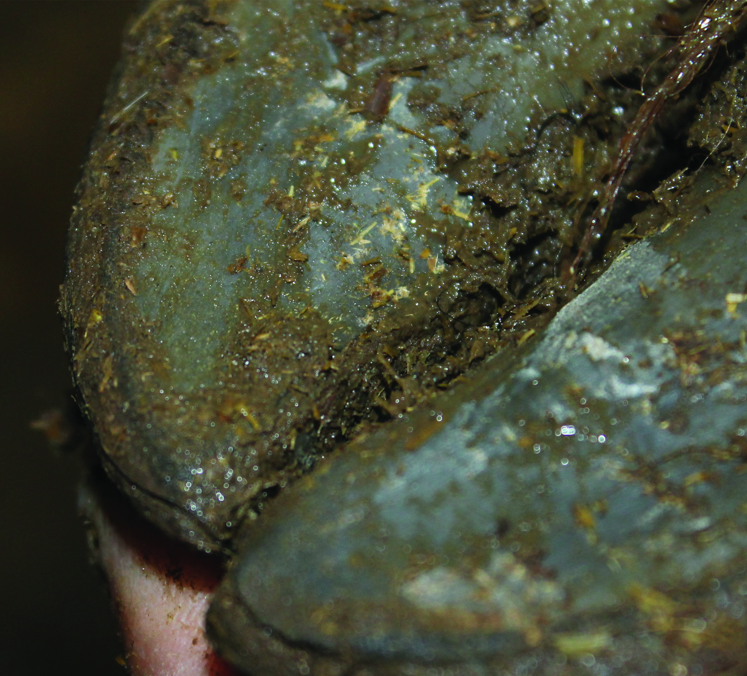
“If you look closely at the foot you see wear at the tip of the toe, and separation along the white line. Depending on which point of the disease you catch it, you might see the start of an abscess or just some separation. In bad cases you’ll find the infection has already penetrated the hoof wall and is going up into P3 (coffin bone) in that claw. The infection can then travel straight up through the tendons and on up the leg and into the bloodstream,” he explains.
Those cattle become septicemic; the pathogens travel to all the organs, and seed into the lungs. “What we see in these animals is a classic embolic pneumonia. When veterinarians see that type of pneumonia—especially in feedlot cattle–they usually associate it with liver abscesses, and the last place they generally think of is the feet. People rarely check the feet to see if there’s a foot infection to explain the pneumonia.”
There are several risk factors in association with toe-tip necrosis. “Dr. Miskimins, a pathologist in South Dakota, described outbreaks in several feedlots in the midwestern U.S. in the 1980’s. He found cases occurring in clusters, in groups of cattle coming in. He began to wonder if high-strung cattle might be more at risk for this disease. We have investigated this in several situations. Dr. Karen Schwartzkopf-Genswein at the Lethbridge Research Center and Dr. Eugene Janzen and myself have collectively investigated a number of outbreaks and it has become clear that high-strung animals are more likely to get this disease.”
Flighty animals are more apt to injure the hind feet, pushing forward in a chute or scrambling on rough concrete surfaces, pushing against one another. “Cattle have so much strength in the hind legs that they put tremendous forces on their toes. At some point they may slip and lose their purchase on the concrete, and slide the foot along it. The concrete is like a rasp and scrapes the toe off,” he says.
“In animals that die with toe-tip necrosis, the white line is thinner than in animals that die of all other causes. The white line is about 4 to 5 millimeters in depth and if the foot suddenly wears off a couple millimeters, the white line probably starts to break down as the animal is walking, and fissures develop. Bacteria enter the soft while line and produce enzymes that break down the white line even more,” he explains.
As the animal walks, repetitive loading and unloading of the foot leads to further breakdown of the white line. “I worked with Dr. J.D. Johnson in the engineering department, taking claws and putting forces on them. When we load the foot that has already suffered damage, the white line starts opening up more, allowing bacteria to get in,” he says.
“Cattle coming onto concrete after a wet summer on pasture seem more prone to this disease, and that makes sense because their feet are softer and more vulnerable to wearing away on abrasive surfaces. It doesn’t happen in pastured cattle. We have created it by management.”
He and his colleagues also did a study looking at nutrition and micronutrients within the hoof wall and sole. “Prairie Diagnostic Services conducted the analytical work for us. We determined that magnesium levels in the horn tissue–hoof wall and sole—were lower in the cattle with toe-tip necrosis than in the controls. We were unable to measure the calcium, however, because it wasn’t in the standard mineral panel, and calcium is a problem in the integrity of horn tissue. Magnesium and calcium relate, therefore if one is low the other is low. I suspect low magnesium could be an indicator for low calcium,” he says.
Thus there may be a nutritional basis to this problem, as well. “This is probably the lesser issue, however. Trauma to the foot would be the biggest factor. Prevention centers on assessing flooring.”
Ideally you need to provide adequate traction so cattle won’t slip and fall, so it’s a fine line; you want enough traction that they don’t get hurt slipping, but you don’t want it so abrasive that it damages the feet. The other important thing is the cattle handling. Handle them in a quiet way, in the best low-stress manner. “There can be other things that play a role. Most diseases are the result of many factors that all come together at just the right time to tip over the animal’s defenses and develop the disease,” Jelinski explains.
With toe-tip necrosis there may be multiple factors–perhaps fractious animals, soft hooves, rough flooring, poor handling practices, etc. and it ends up in disaster. “Once the infection gets past the corium (the inner soft tissue beneath the hoof horn, with blood supply), it gets into the bone. Once it gets to that point it is very difficult to treat. Thus early treatment is important.”
“We are evaluating best practices for treatment. Should we be nipping these toes, treating them like an abscess—to allow drainage—and give antibiotic coverage as well? We are trying to figure out the best way to treat these,” he says. The most important thing, however, is prevention.
Treatment
For treatment of toe-tip necrosis, long-acting antibiotics are generally of use, and successful if you catch it early. “What often happens, however, is separation at the white line before we realize there’s a problem. It’s not until the infection gets to the corium (the ‘quick’) where the nerves and blood are. At that point it becomes painful and the animal is lame. If you catch it and treat it early, there is blood supply, so antibiotics would be in high concentration and treatment would be successful,” says Jelinski.
“Once it gets deeper and into P3, it’s more difficult; the bone remodels. Once it advances we may have to amputate that toe. If veterinarians are going to amputate a toe, they’d better check the other toes as well. Euthanasia may be the best option on some of these animals.”